Protein structure reveals how a malaria parasite imports a wide range of sugars - Nature.com
Most cases of malaria are caused by the protozoan parasite Plasmodium falciparum1. Given that there are more than 400,000 malaria-associated deaths annually, and that P. falciparum is constantly evolving to resist pharmacological therapies, opportunities for developing drugs that target this organism must be continuously explored. A protein called the P. falciparum hexose transporter 1 (PfHT1) has a proclivity for scavenging sugars from an infected host’s red blood cells to improve the parasite’s chances of survival in these cells, and is therefore a drug target. Writing in Nature, Qureshi et al.2 describe the 3D structure of PfHT1, and identify a mechanism that couples the docking of a sugar in the PfHT1 binding site to the process by which sugars are gated through the protein. This coupling facilitates the protein’s substrate promiscuity — that is, its ability to transport a wide range of sugar molecules effectively, a feature that gives the parasite a distinct survival advantage.
Transporter proteins shuttle substrate molecules across the otherwise impermeable lipid bilayer of the cell membrane. The functional and dynamic properties of these membrane-embedded proteins are fundamentally related to their 3D structures, which are modulated at the atomic level over a broad range of timescales. Membrane transporters use the alternating-access mechanism for gating3, in which access to the substrate-binding site switches from one side of the membrane to the other (Fig. 1).

Figure 1 | The alternating-access mechanism. Transporter proteins facilitate the passage of substrate molecules across cell membranes. Access to the substrate-binding site in the middle of transporters is controlled by two gates (red). a, In the outward open state, a pathway from the cell exterior allows substrates into the protein. b, In the outward occluded state, a substrate is trapped between the gates, but the outward-facing pathway is still present. c, In the fully occluded state with a bound substrate, no pathways are available. d, In the inward occluded state, a pathway to the cytoplasm has formed, but the gate remains closed. e, In the inward open state, substrates can exit to the cytoplasm. Qureshi et al.2 report the structure of PfHT1, a sugar transporter from the malaria parasite Plasmodium falciparum. They find that the binding of a sugar substrate to the structure shown in a is coupled to the gating mechanism, and that the transition from a to c occurs much faster than in other sugar transporters. This explains why PfHT1 transports a wide range of sugar molecules equally effectively, unlike other sugar transporters.
The development of methods for determining the structures of membrane proteins in the past few years has produced near-complete pictures of the translocation mechanisms of several classes of transporter — that is, the global rearrangements that the proteins undergo during translocation cycles of substrate binding, transport and release have been visualized at atomic resolution. Intuitively, the substrate specificity of transporters has generally been found to depend on the amino-acid residues at the binding site. The structure of PfHT1 now implies that another mechanism affecting substrate specificity might be at play.
Red blood cells infected by P. falciparum consume about 100 times more glucose than do non-infected cells4 because the parasite continuously metabolizes sugars from these cells to support its growth and replication. Because PfHT1 is responsible for transporting sugars from host cells, it has a crucial role in supporting this metabolism. It belongs to the well-studied major facilitator superfamily (MFS) of transporters, which promote the diffusion of substrates across the cellular membrane. It has the same overall 3D structure as the distantly related human GLUT transporters5. But whereas these specialize in the transport of either d-glucose or d-fructose, PfHT1 transports both of these sugars, and some others, with comparable efficiency.

Qureshi et al. resolved the 3D structure of PfHT1 in which d-glucose is captured in the sugar-binding site, and found that the protein was in a fully occluded conformation — that is, the transporter protein completely shielded the sugar from the aqueous environments on either side of the cell membrane. The structure therefore provides a snapshot of the substrate during a part of the translocation cycle that had not previously been visualized for an MFS transporter.
Armed with their structure, the authors carried out extensive transport studies to try to work out why PfHT1 has less substrate selectivity than its human GLUT counterparts. They first demonstrated that the same set of amino-acid residues in PfHT1 is required to bind d-glucose and d-fructose. They then replaced residues in and around the sugar-binding site of PfHT1 by residues found in GLUT transporters, but none of these mutations conferred GLUT-like selectivity on the resulting proteins. They thus concluded that the unusual lack of selectivity of PfHT1 cannot be explained on the basis of the sugar-binding residues alone.
So how can the substrate promiscuity of PfHT1 be explained? It has been known since the first structures of MFS transporters were reported6,7 in 2003 that bundles of α-helices in the proteins ‘rock’ around the central substrate-binding site, thereby establishing the alternating pathways for substrates through the protein: an outward-facing pathway, which allows substrates into the transporter from the cell exterior, and an inward-facing pathway that allows substrates to enter the cytoplasm (Fig. 1). By considering their structure of the fully occluded state of PfHT1 alongside structures of other sugar transporters captured at different stages in the translocation of d-glucose8–13, Qureshi et al. were able to describe a complete translocation cycle.
The authors found that, surprisingly, all of the sugar-binding residues maintain their orientations throughout the cycle. This implies that the switches from the outward-facing conformation of PfHT1 to the fully occluded state, and then to the inward-facing conformation, are not driven by structural rearrangements at the sugar-binding site. Instead, they are driven by a previously unknown mechanism.
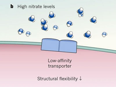
Qureshi and co-workers’ analysis of the gating mechanism of PfHT1 revealed interactions involving hydrophilic amino-acid residues in two transmembrane α-helices in the occluded state. By contrast, in human GLUT proteins, the equivalent residues are larger and more hydrophobic. Experiments in which the authors substituted these gating residues in PfHT1 with other residues demonstrated that they are crucial for sugar transport. Notably, the gating residues are about 15 ångströms away from the sugar-binding site — a large distance. This indicates that the binding of a sugar is coupled to remote conformational changes associated with gating of the transporter, a type of mechanism known as allosteric coupling. Thus, the ability of PfHT1, unlike its human counterparts, to transport many similar substrates results from its substrate-driven gating dynamics, which allows it to adopt the occluded conformation more easily and rapidly.
The authors also carried out experiments to investigate how PfHT1 is inhibited by two small-molecule antimalarial drugs (C3361 and MMV009085). This allowed them to identify a hydrophobic pocket in the transporter that probably facilitates the binding of inhibitory drug molecules, and that might help to guide the design of new antimalarial compounds. However, the most exciting finding is the allosteric coupling between substrate binding and gating — it suggests that substrate recognition in transporters can be a consequence of the transporter’s conformational dynamics, rather than being the result of protein–substrate interactions, which underpin the conventional ‘lock and key’ model of how molecules interact with their biological targets.
References
- 1.
World Health Organization. World Malaria Report 2019 (2019).
- 2.
Qureshi, A. A. et al. Nature https://ift.tt/2RCkRvc (2020).
- 3.
Jardetzky, O. Nature 211, 969–970 (1966).
- 4.
Roth, E. Jr Blood Cells 16, 453–466 (1990).
- 5.
Woodrow, C. J., Burchmore, J. R. & Krishna, S. Proc. Natl Acad. Sci. USA 97, 9931–9936 (2000).
- 6.
Abramson, J. et al. Science 301, 610–615 (2003).
- 7.
Huang, Y., Lemieux, M. J., Song, J., Auer, M. & Wang, D.-N. Science 301, 616–662 (2003).
- 8.
Deng, D. et al. Nature 526, 391–396 (2015).
- 9.
Nomura, N. et al. Nature 526, 397–401 (2015).
- 10.
Deng, D. et al. Nature 510, 121–125 (2014).
- 11.
Sun, L. et al. Nature 490, 361–366 (2012).
- 12.
Quistgaard, E. M., Löw, C., Moberg, P., Trésaugues, L. & Nordlund, P. Nature Struct. Mol. Biol. 20, 766–768 (2013).
- 13.
Wisedchaisri, G., Park, M.-S., Iadanza, M. G., Zheng, H. & Gonen, T. Nature Commun. 5, 4521 (2014).
Latest on:
Molecular biology
Structural biology


Comments
Post a Comment